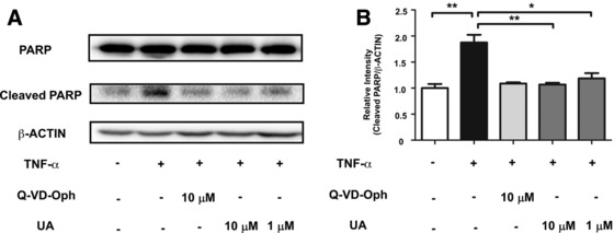
Figure 4.

UA attenuated TNF‐α‐induced CASP3 activation and proteolytic processing of PARP (A) CHO cells were stimulated by 20 ng mL−1 TNF‐α for 12 h with or without 6 h pretreatment with UA or Q‐VD‐Oph. The cleaved PARP was measured by Western blot analysis. (B) The relative intensity data of cleaved PARP to β‐actin represent mean ± SD of three group; *p < 0.05 and **p <0.01.
