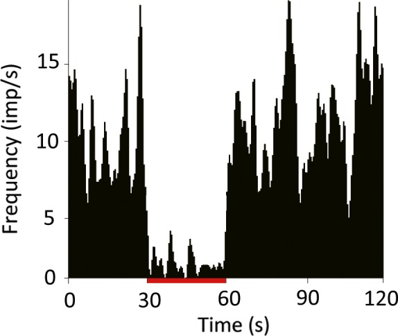
Figure 5.

Changes in GPi‐neuron activity in response to STN‐HFS. The spiking activity of most globus pallidus pars interna (GPi) neurons (82%) was reduced by STN‐HFS, while a small minority (12%) showed an increase in excitability (spike rate) (P < 0.05 vs. baseline spike rate, Mann–Whitney U‐test). Red bar indicates stimulation period (30 s). [Colour figure can be viewed at wileyonlinelibrary.com].
