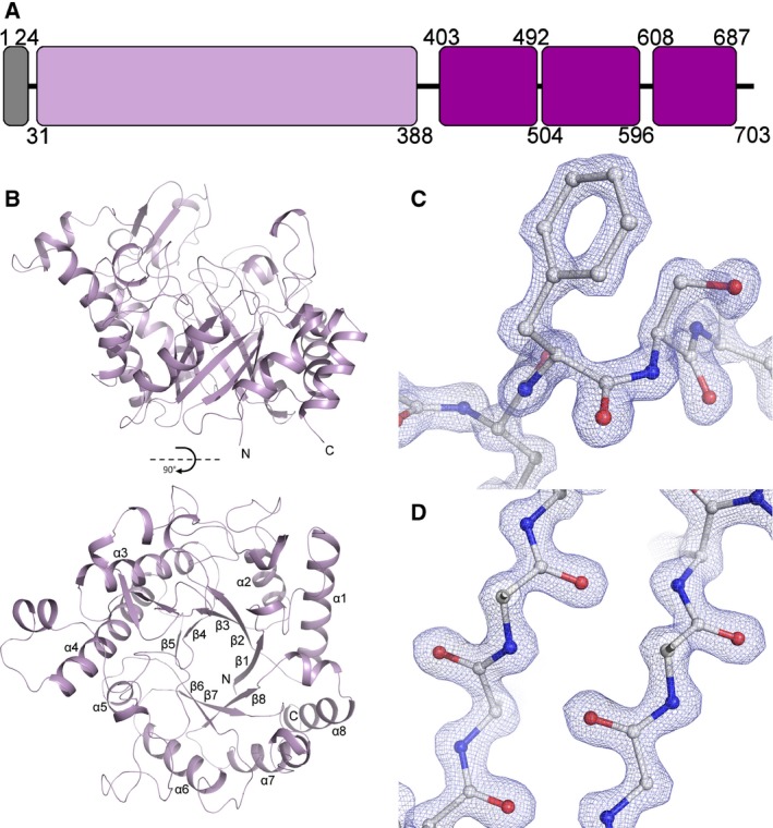
Figure 1.

The Structure of Cwp19‐fr. (A) Domain representation, the signal peptide is coloured grey, the GHL10 domain is lilac and the cell wall‐binding domains are purple. The present construct codes for residues 27‐401, while residues 28‐388 are visible in the structures. (B) Ribbon diagram of the overall fold. Cwp19‐fr assumes a TIM barrel fold, forming an eight‐stranded β‐barrel surrounded by eight α‐helices. The active site is formed over the centre of the barrel near the C‐termini of β‐strands. The two images are related by a 90° rotation on the x‐axis. (C) Example electron density, 2FO‐FC, 2.5σ. The cis‐peptide formed by Phe367 and Ser368 is shown. (D) Sections of β‐strands 5 and 6.
