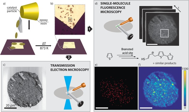
Figure 1.

Integrated SMF microscopy and TEM of a single catalyst particle. a) Fluid catalytic cracking (FCC) particles embedded in epoxy resin (yellow) are microtomed into thin sections and deposited onto a SiN membrane. b) Calcination of the SiN membrane removes the resin, leaving just the catalyst thin sections. c) TEM image of the thin section. d) Sample reactivity is evaluated by SMF using the thiophene oligomerization as probe reaction; a movie with 9200 frames is recorded (Movie S1), showing the emitted fluorescence as bright, diffraction‐limited spots. The movie is analyzed by NASCA (e) and SOFI (f). e) Map of detected single‐molecule events by NASCA. For clarity, the detected events have been enlarged. Fewer events are observed in the top right area because it is slightly out of focus. f) Map of the SOFI intensity. The scale bars represent 20 μm.
