
Figure 4.
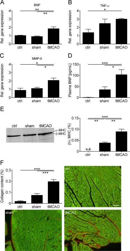
Increase of biomarkers for cardiac dysfunction and slow left ventricular (LV) remodeling after right transient middle cerebral artery occlusion (tMCAO). (A–C) Relative (Rel.) gene expression of brain natriuretic peptide (BNP) (A), tumor necrosis factor α (TNF‐α) (B), and matrix metallopeptidase‐9 (MMP‐9) (C) in the LV tissue of control (ctrl), sham‐operated, and tMCAO‐treated mice 8 weeks after surgery (n = 4 or 5 per group; *p < 0.05, **p < 0.01). (D) Plasma BNP levels from ctrl, sham‐operated, and tMCAO‐treated mice 8 weeks after tMCAO (n = 8–12 per group; *p < 0.05, ***p < 0.001). (E) Left panel: representative silver‐stained sodium dodecyl sulfate–polyacrylamide gel electrophoresis of α‐ and β‐myosin heavy chain (MHC) distribution in the left ventricle of ctrl, sham‐operated, and tMCAO‐treated mice 8 weeks after surgery. Right panel: densitometric quantification (n = 4 per group; **p < 0.01, ***p < 0.001; n.d. = not detected). (F) Densitometric quantification and representative images of the collagen (red) content in LV tissue (green) sections (7 µm) using picrosirius red staining (n = 4 or 5 per group; scale bar = 50 µm; ***p < 0.001).