
Figure 7.
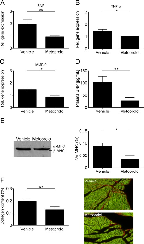
Influence of metoprolol on the cardiac biomarker profile and left ventricular (LV) remodeling. (A–C) Relative (Rel.) gene expression of brain natriuretic peptide (BNP) (A), tumor necrosis factor α (TNF‐α) (B), and matrix metallopeptidase‐9 (MMP‐9) (C) in the LV tissue of vehicle‐ and metoprolol‐treated mice 8 weeks after surgery (n = 5 per group; *p < 0.05, **p < 0.01). (D) Plasma BNP levels from vehicle‐ and metoprolol‐treated mice 8 weeks after transient middle cerebral artery occlusion (n = 8–11 per group; **p < 0.01). (E) Left panel: representative silver‐stained sodium dodecyl sulfate–polyacrylamide gel electrophoresis of α‐ and β‐myosin heavy chain (MHC) distribution in the left ventricle of vehicle‐ and metoprolol‐treated mice 8 weeks after surgery. Right panel: densitometric quantification (n = 4 per group; *p < 0.05). (F) Densitometric quantification (left panel) and representative images (right panel) of the collagen (red) content in LV tissue (green) sections (7 µm) using picrosirius red staining (n = 4 or 5 animals per group; scale bar = 50 µm; **p < 0.01).