Brain MRI
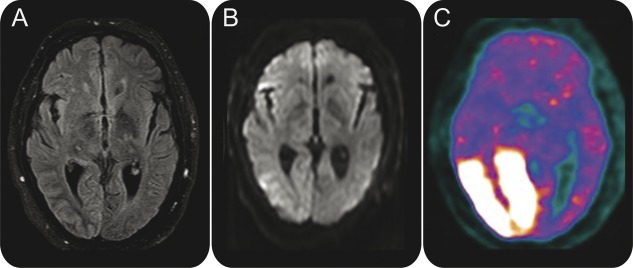
Figure 3. Brain images of patient 2 demonstrate right occipital cortical hyperintensity and subcortical hypointensity on fluid-attenuated inversion recovery (A), subtle restricted diffusion in lateral right occipital lobe on diffusion-weighted imaging (B), and marked hypermetabolism of right occipital lobe on FDG-PET (C).

