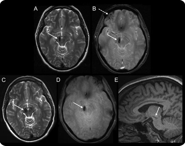
Head MRI
Figure. (A) Axial T2-weighted sequence shows focal hypointensity (arrow) in the right ventral midbrain just lateral to the red nucleus and extending rostrally into the substantia nigra. (B) Axial T2* gradient echo sequence shows focal hypointensity in the right midbrain (solid arrow) and right frontal lobe (dashed arrow). There are similar hypointensities involving the left frontal lobe. These susceptibility artifacts are indicative of hemorrhagic lesions. (A, B) About 5 months after head trauma. (C–E) Axial T2-weighted, axial T2* gradient echo, and sagittal T1-weighted sequences that were obtained about 8 years after head trauma. (C, D) No appreciable evolvement of the right midbrain lesion. (E) The rostro-caudal extent of the lesion and its involvement of the substantia nigra.
