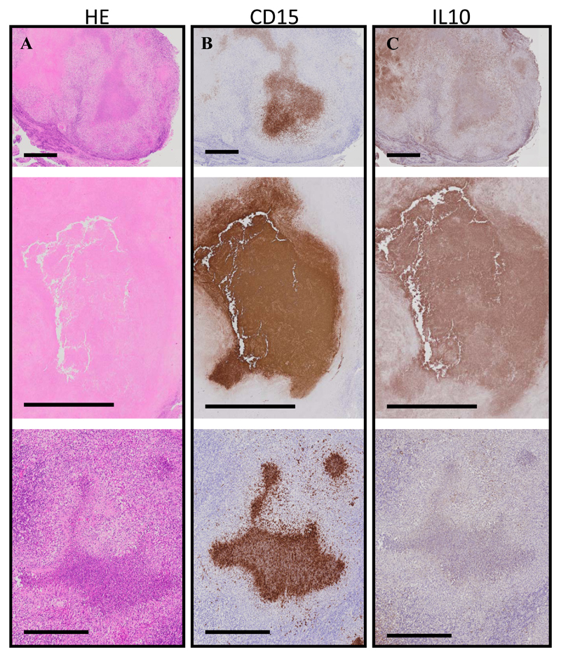
Figure 5. Neutrophil infiltration in the lymph nodes of TB-IRIS patients.
Caseous granulomas from consecutive cross-sectional lymph node sections of TB-IRIS patients (n = 3) that were stained with Hematoxylin and Eosin (H&E) (A), CD15 (neutrophils, B), or IL-10 (C). Intense neutrophil staining localizes within most of these caseous granulomas. IL-10 staining was diffuse but did localize within and near caseous granulomas. Black bars represent 200 μm.

