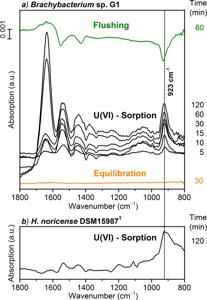
Fig 6.
a) In situ ATR FT-IR difference spectra of U(VI) sorption on Brachybacterium sp. G1 cells ([U(VI)] = 40 μM, pCH+ 6, 1.7 M NaCl). The “Equilibration” spectrum confirms a stable bacterial film on the ATR crystal. “U(VI)—sorption” spectra were recorded at different times after induction of U(VI) association. “Flushing” shows the reversibility. b) For comparison the spectrum of U(VI) bioassociation on H. noricense cells after 120 min ([U(VI)] = 40 μM, pCH+ 6, 3 M NaCl) [11].

