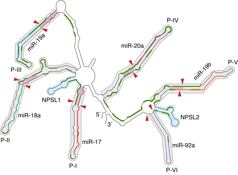
Figure 3.
Schematic of the structural map showing domain architecture in oncomiR-1. OncomiR-1 is empirically divided into helical domains, P-I to P-VI (grey shaded regions), containing individual pri-miRNA helices and two non-pri-miRNA stem loop domains NPSL1 and NPSL2 (cyan shaded regions). The regions corresponding to mature miRNA sequences in P-I to P-VI are indicated as colored segments (e.g. miR-17 is in red, miR-19a in blue, miR-19b in orange etc.). Red arrowheads indicate Drosha cleavage sites on oncomiR-1. The pink shaded regions indicate the basal helix of each pri-miRNA domain. Solvent inaccessible segments on oncomiR-1 are highlighted in green.

