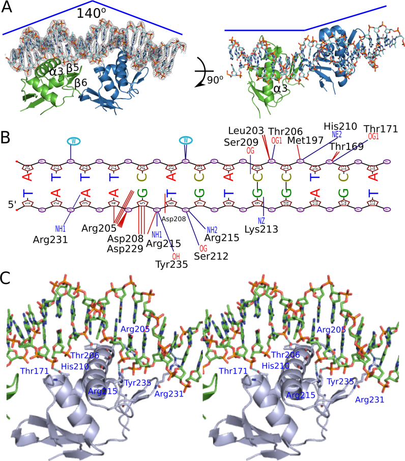
Figure 4.
Crystal structure of AdeR DNA binding domain in complex with the intercistronic DNA. (A) Overall structure of AdeR_DBD with DNA complex. Two monomers of AdeR_DBD binds with the two direct repeat intercistronic regions of DNA. The α3 and β hairpin formed by the β5 and β6 are involved in the binding of major and minor groove respectively. The density of the DNA in the left panel is shown in 2Fo – Fc = 1.5σ. (B) NUCPLOT of the detailed interaction between one AdeR_DBD and a single repeat of the intercistronic DNA. The dark blue represents the hydrogen bonds. (C) Detailed main interaction between the AdeR DNA binding domain and the intercistronic DNA.

