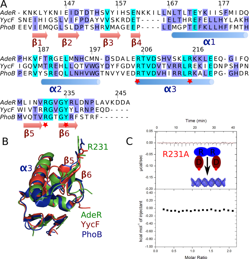
Figure 5.
Sequence alignment of AdeR DNA binding domain and mutagenesis study. (A) Sequence alignment of AdeR with the OmpR/PhoB like response regulator YycF and PhoB DNA binding domains. The red star indicates the key positive residues involved in the protein and DNA interaction. (B) Superposition of AdeR (Green) with YycF (Red) and PhoB (Blue) DNA binding domains with RMSD of 0.8 and 1.0 Å respectively; R231 is shown in stick. (C) The R231A mutation in AdeR full length could abolish its interaction with the intercistronic DNA as shown by isothermal titration calorimetery.

