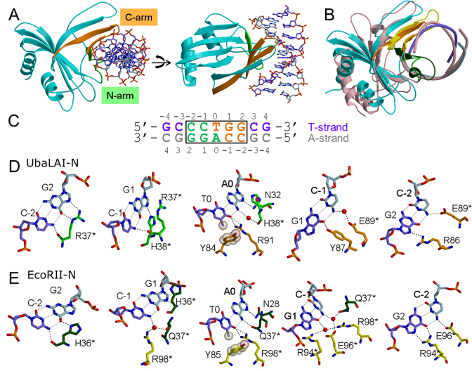
Figure 4.
UbaLAI-N–DNA complex structure. (A) Two different views of UbaLAI-N–DNA complex structure. The secondary structure elements of the N-arm are shown in green; those belonging to the C-arm are shown in orange, the T-strand of DNA is shown in violet. (B) Superposition of UbaLAI-N and EcoRII-N DNA complexes. UbaLAI-N structure is colored as in (A), EcoRII-N–DNA complex is colored pink. N-arm and C-arm of EcoRII-N are colored dark green and yellow, respectively. (C) Oligonucleotide used for crystallization of UbaLAI-N. Recognition sequence is boxed, DNA bases recognized by residues from N-arm and C-arm are shown in green and orange letters, respectively. (D) Recognition of the 5′-CCTGG-3′ sequence by UbaLAI-N. Panels for individual base pairs are arranged following the T-strand in 5′-3′ direction. Residues belonging to the N- and C-arms are colored in green and orange, respectively. (E) Recognition of the 5′-CCTGG-3′ sequence by EcoRII-N. Residues belonging to the N- and C-arms are colored in dark green and yellow, respectively.

