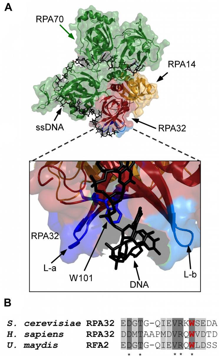
Figure 1.

Position of non-canonical amino acid insertion in RPA. (A) Crystal structure of Ustilago maydis RPA bound to ssDNA is shown (PDB ID: 4GOP) with RPA70, RPA32 and RPA14 colored green, red and yellow, respectively. The zoomed-in image shows two loops, L-a and L-b, flanking the ssDNA (black sticks) and Trp-101 is shown as stick representation in blue. (B) Conservation of amino acid sequence in the region where Trp-101 resides in RPA32. W101 is highlighted in bold (red).
