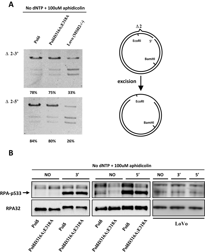
Figure 2.
Mismatch-provoked excision in HeLa cells expressing PolδD316A;E318A. (A) Mismatch provoked excision assay was performed as described in Materials and Methods using the nuclear extracts indicated. The extent of DNA excision was estimated by measuring susceptibility/resistance to cleavage by EcoRI, whose recognition sequence lies in between the DNA excision initiation site and the mismatch (schematic diagram right). Reaction products were digested with EcoRI and BamHI, separated by agarose gel electrophoresis and visualized by staining with ethidium bromide. The intensity of each band was quantified using ImageJ, and the relative excision capacity was calculated as the intensity of the slowest migrating (largest) reaction product per lane /total intensity per lane × 100. Nuclear extracts from MSH2-deficient LoVo cells were used as the negative control. (B) Western blot of pS33-RPA32 was performed to monitor amount of ssDNA generated during in vitro MMR. Briefly, MMR assay was performed as described in Materials and Methods with no substrate (NO), Δ2–3′ (3′) or Δ2–5′ (5′) substrate in the reaction. The MMR was terminated by adding SDS containing loading buffer and boiling at 95°C for 10 min. Western blot was performed as described in Materials and Methods. Aphidicolin was included and dNTPs were omitted as indicated for inhibition of polymerase synthesis of Polδ. The total RPA32 was used as control.

