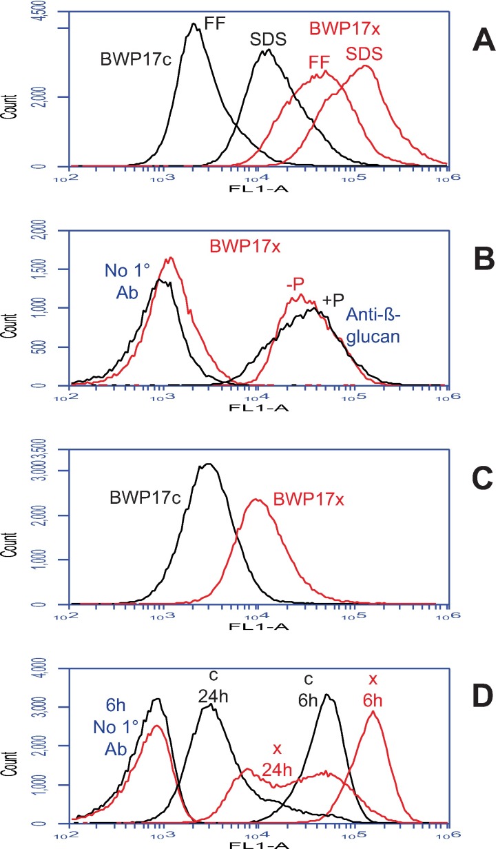
Fig 4. Strain BWP17x also binds more anti-β-1,3-glucan.
Flow cytometric analysis of parental (BWP17c) and mutant (BWP17x) yeast forms after labeling with a monoclonal mouse anti-β-1,3-glucan IgG followed by fluorescein-conjugated goat anti-mouse IgG. Cells were cultured as in Fig 2. (A) Relative to formaldehyde fixation (FF), extraction with SDS at 70°C enhanced anti-β-1,3-glucan binding to both BWP17c and BWP17x, but BWP17x showed far more binding than BWP17c after both treatments. (B) Phosphate limitation had little effect on anti-β-1,3-glucan binding; also shown in this panel are controls that omitted the primary antibody (1° Ab) but included the secondary (fluorescein-conjugated) antibody. (C) Replacement of glucose with lactate as the sole carbon source in the growth medium diminished anti-β-1,3-glucan binding to BWP17x, but to a level that was still almost 4× that of BWP17c. (D) Cells in early phase cultures (6 hr in YPD) showed more binding of anti-β-1,3-glucan than cells at later phases (shown for 24 hr); the later phases showed much more variability, however, based on the width of the profiles. Populations in panels B, C and D were gated as in Fig 2 to exclude aggregates and multiplets.

