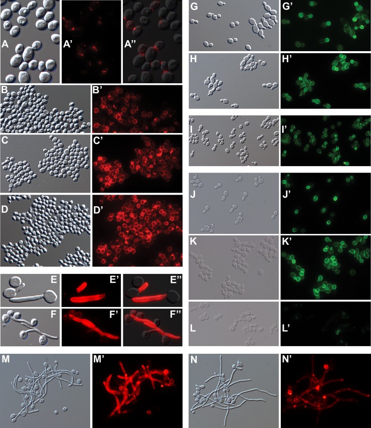
Fig 5. Micrographs of cells labeled with anti-β-1,3-glucan.
DIC (A-N) and corresponding fluorescence (A’-N’) micrographs. The secondary antibody was goat anti-mouse IgG conjugated to either AlexaFluor 594 (A’-D’, M’, N’) or fluorescein (G’-L’). (A”, E”, F”) Composite overlay of DIC and fluorescence images. BWP17c (A,B) and BWP17x (C,D) grown as in Fig 3C and 3D without (A,C) or with (B,D) Caspofungin. BWP17c (E) and BWP17x (F) after 3 hr in 37°C RPMI filamentation medium. BWP17c (G) and BWP17x (H) after 6h in 30°C YPD medium. (I) BWP17x after extraction with SDS at 70°C. Ywp1-knockout strain 4L1 after growth in BMM13 for 4h (J), 8h (K) or 14h (L). Strains devoid of Ywp1 (M) or ectopically producing Ywp1 in germ tubes and hyphae (N) after 3.5 hr in 37°C RPMI filamentation medium. Actual width of each image (μm): A: 27; B: 93; C: 97; D: 101; E: 30; F: 43; G: 83; H: 94; I: 132; J: 107; K: 91; L: 118; M, N: 102.

