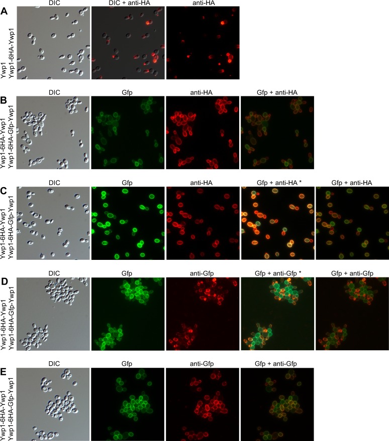
Fig 14. Detection and localization of Gfp and HA tags in Ywp1 (part 1).
Micrographs in each row show corresponding DIC images, intrinsic Gfp fluorescence (Gfp), antibody-amplified epitope fluorescence (red anti-HA and anti-Gfp), and digital overlays of the green and red images; in some cases (*), both the red and green fluorescence were photographically captured in the same image. The actual width of each image is 88 μm. Cells were grown in 30°C BMM13 for 20 hr, then transferred to 30°C BMM13 lacking phosphate for 5 hr (A,B,D) or 23 hr (C,E) prior to formaldehyde fixation and labeling with monoclonal primary antibody (anti-HA or anti-Gfp) and AlexaFluor 568 conjugated secondary antibody. The versions of Ywp1 present in each strain are shown at the left of each row; row A is strain YHY and rows B-E are a derivative of strain YHYx.

