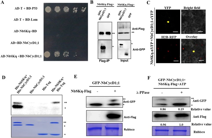
Fig 6. NbSKη interacts with and phosphorylates NbCycD1;1.
(A-C) Interaction between NbCycD1;1 and NbSKη was validated in Y2H (A), Co-IP (B) and BiFC (C) assays. ** indicates NbSKη-Flag. * represents the substrate of NbSKη. (D) NbSKη phosphorylates NbCycD1;1 in vitro. The upper panel shows autoradiography and the bottom panel shows coomassie blue staining. ** indicates His-NbSKη. * represents the substrate of NbSKη. (E) NbSKη phosphorylates NbCycD1;1 in vivo. ** indicates phosphorylated form of GFP-CycD1;1. * represents the non-phosphorylated form of GFP-CycD1;1. (F) Identification of the phosphorylation of NbCycD1;1 mediated by NbSKη using λ-PPase. Plant tissues expressing GFP-NbCycD1;1 with or without Flag-NbSKη were extracted at 60 hpi. Two aliquots of samples were treated in the reaction system in the absence or presence of λ-PPase for only 10 min at 30°C. ** indicates phosphorylated form of GFP-CycD1;1. * represents the non-phosphorylated form of GFP-CycD1;1. Relative values represent the ratio of phosphorylated form and non-phosphorylated form of NbCycD1;1 (top panel) and the accumulation level of NbSKη-Flag (middle panel), respectively.

