
Figure 1. Postnatal brain maturation is accompanied by a progressive decrease in nociceptin expression.
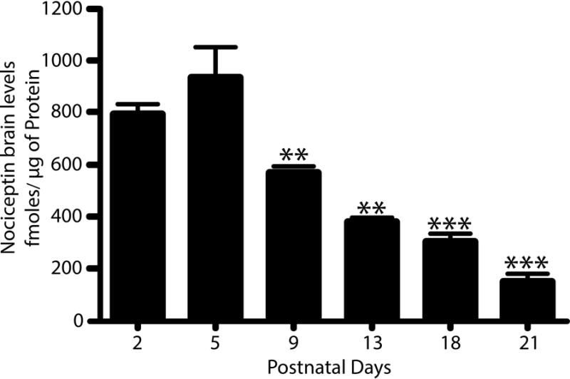
Nociceptin levels were measured by dot blot analysis of total homogenates prepared from cerebral hemispheres of rat pups at 2-, 5-, 9-, 13-, 18-, and 21- postnatal days. Purified nociceptin was used to generate a standard curve as indicated under “Methods”. The results, expressed as fmoles of nociceptin/μg of protein, are the average ± SEM from at least 3 animals per age, PD5 vs. PD2, not significant (n.s.); PD5 vs. PD9 and PD13, **p<0.01; PD5 vs. PD18 and PD21, ***p<0.001.