
Figure 6. Treatment of astrocytes with nociceptin results in a dose-dependent increase in GLAST expression.
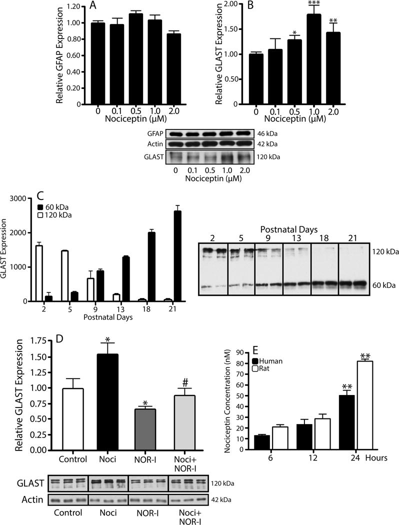
Astrocytes isolated from 3-day-old rat brains were treated for 24 hrs with increasing concentrations of nociceptin. (A) GFAP and (B) GLAST expression were evaluated by western blot analysis, using β-actin as loading control. Results are expressed as change relative to control values and represent the mean ± SEM, n=3, *p<0.05, **p<0.01, ***p<0.001. (C) GLAST levels in total brain homogenates from 2-, 5-, 9-, 13-, 18-, and 21-day-old rats were determined using western blot. Each gel lane was loaded with 5μg of protein and levels for the 60 kDa (black bars) and 120 kDa (open bars) molecular forms of GLAST correspond to the mean ± SEM from 3 animals per age. (D) Western blot analysis was used to determine relative GLAST levels after a 24 hr incubation in CDM alone, 1μM nociceptin (Noci), 100nM BAN-ORL24 (NOR-I), or 1μM Noci + 100nM NOR-I. The results are expressed as change relative to control values and represent the mean ± SEM from at least 3 experiments; control vs. Noci and control vs. NOR-I, *p<0.05; Noci vs. Noci + NOR-I, #p<0.03. (E) Secretion of endogenous nociceptin was assessed by dot blot analysis of medium collected from cultured rat (open bars) and human (black bars) astrocytes after 6, 12, and 24 hrs in CDM alone. The results are the mean ± SEM from 6 different cultures. Rat, 6 hrs and 12 hrs vs. 24 hrs, **p<0.01. Human, 6 hrs vs. 24 hrs, **p<0.01.