
Figure 8. 3H-Aspartate uptake increases in primary rat astrocytes treated with nociceptin.
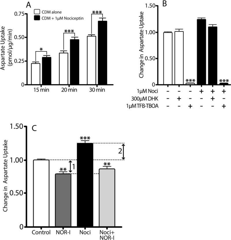
(A) Aspartate uptake in primary rat astrocytes after 24 hr pre-incubation in CDM alone (controls, open bars) or CDM with 1μM nociceptin (black bars) was determined using a 3H-D-aspartate uptake assay, as described under Methods. Results expressed as pmol/μg protein/min and are the mean ± SEM from at least 10 replicates/condition, *p<0.05, ***p<0.001. (B) Aspartate uptake was assessed in cell cultures after 24 hours pre-treatment in CDM alone (white bars) or CDM supplemented with 1μM nociceptin (black bars). Prior to the assay, parallel controls and nociceptin-treated cultures were in addition pre-incubated for 10 min with either 300μM DHK or 1μM TFB-TBOA. Results are expressed as change in uptake over 30 min and represent the mean ± SEM from at least six replicates/condition, control vs control with TFB-TBOA and nociceptin vs. nociceptin with TFB-TBOA, ***p<0.0001. (C) Aspartate uptake was assessed in primary rat cultures treated for 24 hrs with CDM alone (control), or CDM supplemented with either 100nM NOR-I, 1μM nociceptin, or 1μM nociceptin with 100nM NOR-I. Results are expressed as change in uptake over 30 min and represent the mean ± SEM from at least six replicates/condition. Control vs. NOR-I, **p<0.01; control vs. nociceptin, ***p<0.001; nociceptin vs. nociceptin with NOR-I, ***p<0.001. Arrow 1: aspartate uptake due to endogenously produced nociceptin. Arrow 2: aspartate uptake due to exogenously added nociceptin.