
Figure 9. In vivo treatment with an NOR inhibitor or genetic ablation of NOR affect GLAST brain expression.
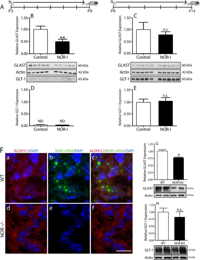
(A) Rat pups were given daily IP injections of BAN-ORL24 (NOR-I) on two different timelines: from postnatal day 3 to 9, or postnatal day 9 to 14. Total homogenates prepared from brains collected at the end of each timeline were subjected to western blot analysis for GLAST and GLT-1. (B and C) GLAST expression in total brain homogenates of animals injected from (B) postnatal day 3 to 9 and (C) postnatal day 9 to 14. (D and E) GLT-1 levels in brain homogenates from pups injected from (D) postnatal day 3 to 9 and (E) postnatal day 9 to 14. The results are expressed as change relative to control values and represent the mean ± SEM from at least 5 animals. **p<0.01; n.s., not significant; ND, not detected. (F) 5-day-old WT and NOR KO mouse cortical brain tissue slices were subjected to immunohistochemical staining with (a and d) anti-ALDH1L1 antibody and in situ hybridization using (b and e) a digoxigenin-labeled probe for NOR mRNA, as indicated under “Methods”. Notice the presence of NOR mRNA in ALDH1L1-labeled astrocytes in WT mice (a-c) and lack of NOR mRNA in the NOR KO animals (d-f). Scale bar: 10μm. (G and H) Total brain homogenates from 14-day-old WT and NOR knockout mice were subjected to western blot analysis to evaluate expression of (G) GLAST and (H) GLT-1, using β-actin as loading control. Results are represented as change relative to WT and are the mean ± SEM from 4 animals, * p<0.01.