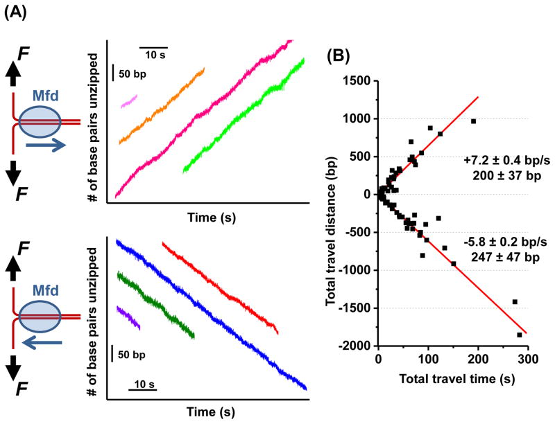
Figure 1. Mfd translocates along DNA on its own.
(A) The DNA “unzipping tracker” assay was used to monitor the translocation of Mfd away from the fork (top panel) and towards the fork (bottom panel) in real time under a constant unzipping force of 18 pN in 2 mM ATP. Multiple real-time tracking traces from different Mfd molecules are shown, with their time and location axes arbitrarily shifted. Each trace starts when the unzipping fork encountered an Mfd and ends when the Mfd dissociated from the DNA.
(B) A scatter plot of all traces for both translocation directions (N = 101). Each trace yields a single data point on this plot, with total distance of travel plotted against total time of travel. The means and standard errors of the means are shown for each direction. See also Figure S1 and Table S2.

