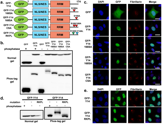
Figure 2.
(a) Structure of GFP-tagged Y14 serine-alanine substitution mutants and the RKPL mutant, which mimics dephosphorylated Y14. (b) Expression of transfected GFP-Y14 and serine-alanine substitution mutants was detected by western blotting with an anti-GFP antibody (upper panel). Phosphorylation status of Y14 and mutants was analyzed by separation using phos-tag gels (bottom panel). The samples for SDS-PAGE and phos-tag SDS-PAGE derive from the same experiment. The cropped blots are used in the figure. The membranes were cut prior to exposure so that only the portion of gel containing the desired bands would be visualized. Full-length blots are shown in Supplementary Fig. S10. (c) Localization of GFP-Y14 and its associated mutants was observed by fluorescence microscopy (green). Fibrillarin and nuclear DNA are shown as red and blue, respectively. Bar indicates 20 μm. (d) Expression of transfected GFP-Y14 and RKPL mutant was detected by western blotting with an anti-GFP antibody (left panel). Phosphorylation status of Y14 and mutants was analyzed by separation using phos-tag gels (right panel). (e) Localization of GFP-Y14 and the RKPL mutant was observed by fluorescence microscopy (green). Fibrillarin and nuclear DNA are shown as red and blue, respectively. Bar indicates 20 μm.

