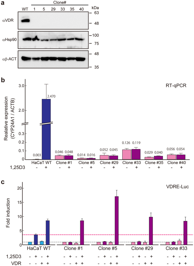
Figure 4.
Confirmation of loss-of-function of the VDR gene in the knocked-in cell lines. (a) Immunoblot analyses of isolated clones. αHsp90 and αβ-ACT were used as loading controls. (b) RT-qPCR analysis of CYP24A1 of the isolated cells with or without treatment with 1α,25-dihydroxyvitamin D3 (1,25D3). (c) Promoter-reporter assay using vitamin D response element (VDRE). When endogenous VDR was functional, VDRE would respond to 1,25D3 treatment. The exogenous VDR expression plasmid (VDR+) was transfected for the complementation assay.

