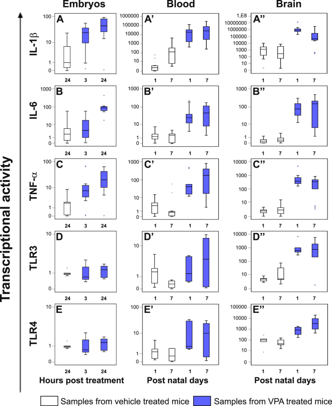
Figure 4.
Expression levels of IL-1β, IL-6, TNF-α, TLR3 and TLR4 genes in whole embryos and, from the offspring, in blood and brain samples from VPA- and vehicle- treated CD-1 dams. The cytokines and TLRs expression in whole embryos (panel A–E) explanted 3 and 24 hours post the treatment, in blood (panel A’–E’) and brain (panel A”–E”) samples obtained at post natal day 1 and 7 from VPA- and vehicle- treated CD-1 dams, was evaluated by real time-PCR and represented as box plots, depicting mild (black dot) and extreme (asterisk) outliers for each group. Prenatal exposure to VPA at gestational day 10.5 increased cytokines expression in embryos (blue box plots), already from 3 hours post treatment than in controls (white box plots). In blood and in brain, also the expression levels of TLRs were higher in VPA mice (blue box plots) than in controls (white box plots).

