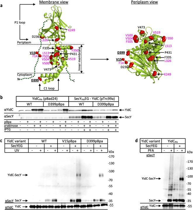
Figure 1.
YidC contacts the SecYEG translocon via TM1 and the C1-loop in vivo. (a) The crystal structure of E. coli YidC (PDB accession no.: 3WFV) visualised from the membrane (left) or from the periplasmic side (right). The red spheres indicate the positions for pBpa insertion. Residues which show the strongest contacts to SecY are displayed in bold and underlined. Residues with weaker contacts are shown in black and residues that did not show significant cross-links are shown in magenta. The dashed green lines indicate TM1 and the C-terminus of YidC, which have not been crystallized so far. (b) The co-expression system shows balanced expression of YidC and SecYEG as revealed by western blotting. 1 × 10⁸ E. coli BL21 cells expressing yidC under the arabinose promoter from plasmid pBad24 or co-expressing PlacsecYEG-yidC from plasmid pTrc99a were TCA precipitated, separated by SDS-PAGE and after western transfer decorated with the indicated antibodies. WT refers to wild type YidC and D399pBpa to a YidC variant with pBpa inserted at position 399, when pBpa was added to the growth medium. (c) In vivo photo-cross-linking performed with BL21 E coli cells expressing either yidC alone (-SecYEG) or co-expressing yidC and secYEG (+SecYEG). Either wild type YidC (WT) or YidC variants containing pBpa at position V15 or D399 were analysed. After UV-exposure, samples were purified via metal affinity chromatography using an N-terminal His-tag on YidC. A sample without UV-exposure served as a control. Samples were decorated with antibodies against SecY or YidC as indicated. The 95 kDa YidC-SecY cross-link is indicated. (d) In vivo para-formaldehyde (PFA) cross-linking with BL21 cells expressing either only YidC from plasmid pBad24 or YidC together with SecYEG from plasmid pTrc99a. All samples were treated as described in experimental procedures and YidC was further purified from the membrane fraction via its N-terminal His-tag and analyzed by western blotting using α-SecY antibodies for detecting SecY-YidC cross-links. α-YidC antibodies determined comparable amounts of YidC in all samples. Uncropped images are displayed in Supplementary Figure S4.

