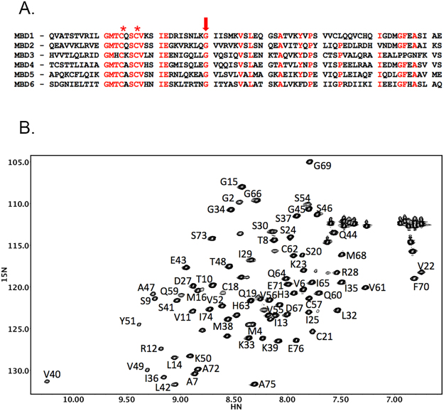
Figure 2.
Amino acid sequence of the metal binding domains of ATP7B (A) and a fingerprint 1H,15N-HSQC spectrum of MBD1 (B). (A) Conserved residues are shown in red. Cysteine residues in the copper binding motif of the MBDs are marked by an asterisk. The invariant glycine, which is a target of the Wilson disease causing mutation in MBD1 (G85V), is shown by the arrow. (B) The sequential amino acid assignments in MBD1 are shown. In the protein construct used for structure determination, residues 1–4 are from the purification tag, and Q5 corresponds to Q56 in the full length ATP7B.

