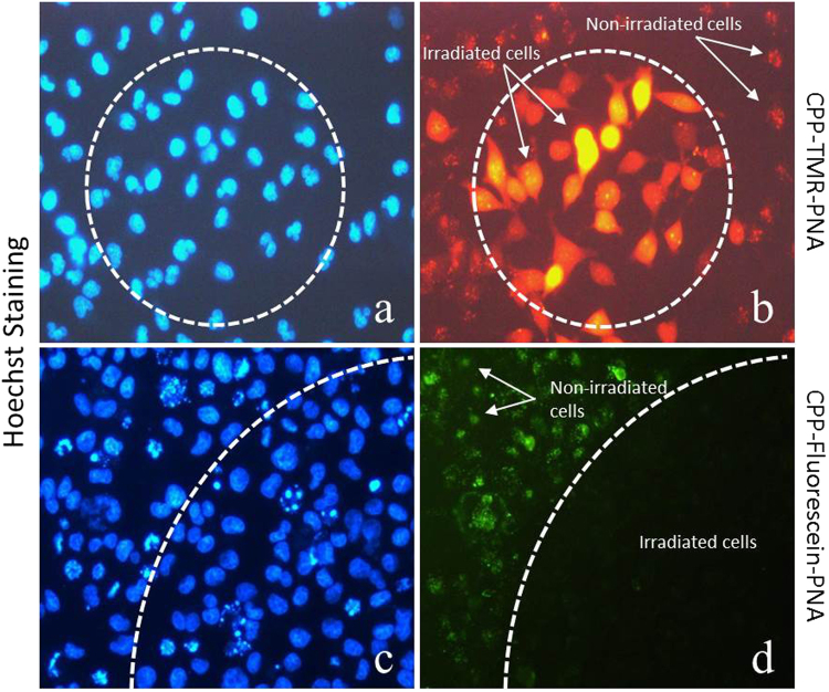
Figure 6.
Controlled site specific effect of PCI. Cells were transfected with 3 µM PNA. (a,c) Cell nuclei were stained with Hoechst for 15 min.; (b) Cells transfected with TMR conjugated PNA irradiated at 555 nm; (d) Cells transfected with fluorescein conjugated PNA irradiated at 490 nm. Irradiation was confined to the dotted areas (dotted lines). Fluorescence microscope (Diaplan, leitz) with 20× objective lens was used for irradiation and imaging.

