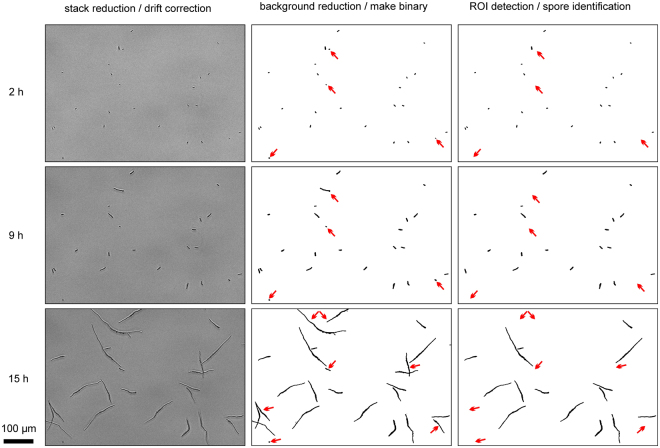
Figure 3.
Image processing by HyphaTracker macro for automated data evaluation. Germination of conidia of the CarO-deficient F. fujikuroi strain was recorded with a frame rate of 0.2 min−1 in a 16-bit image time series (HT-teststack1). Three different time points as indicated are given to visualize the processing procedure of the images by HyphaTracker. In the left column 16-bit images are shown after data reduction and drift correction. The hyphae are slightly out of focus to enable dark appearance in the microscope image. In the middle column the result of step 3 (Binary image generation) of HyphaTracker is shown. In step 4 and 5 (right column) the ROIs are identified and filtered for their size and eventually occurring crossing events or contact to the edge. Red arrows highlight such germlings that were automatically removed from the analysis. Black bar represents 100 µm.

