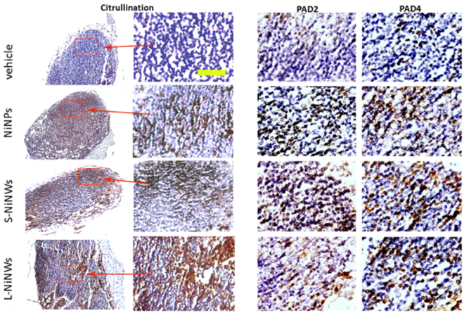
Figure 7.
Immunohistochemistry of anti-citrulline, anti-PAD2 and PAD4 antibodies on murine lymph node (LN) tissue sections. Immunodetection of citrullinated proteins, PAD2 and PAD4 enzymes in murine LN tissue sections 2-week post treatment with nickel nanomaterials and vehicle controls. Brown staining demonstrates positive immunohistochemical reaction for citrulline, PAD2 and PAD4. First two panels from left (Citrulline) represent the corresponding microscopic fields at lower (10×) and higher (20×) magnification stained for citrullinated proteins, to illustrate the topography of positive staining within the LN. Red rectangles indicated by arrows outline the regions of interest magnified in the adjacent panel. Two panels on the right show representative topographically similar, but not identical microscopic fields stained for PAD2 and PAD4 enzymes, respectively. Scale bar (yellow): 100 μm.

