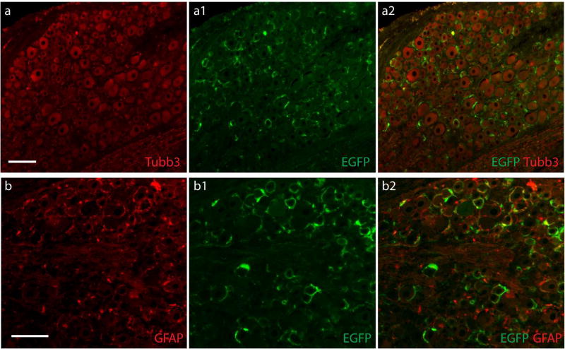
Figure 6. SGC transduction after intraganglionic injection of AAVshH10-GFAP-EGFP.
Representative images from DRGs 5 weeks after injection of 2.0×l010 GC per DRG of AAVshH10-GFAP-EGFP. Data are representative of 6 DRGs from three rats. Efficient transduction was evident by EGFP expression in SGCs clustered in ring-like arrangements (a1) around the majority of DRG neurons labeled by the pan-neuron maker Tubb3 (a, a2). Double labeling with GFAP (b) demonstrates clear overlay of EGFP (b1) with the SGCs labeled by GFAP (b, b2). Scale bar: 100µm for all panels.

