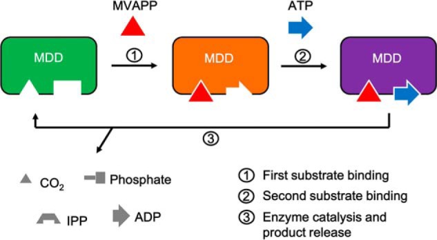
Figure 7.

Induced substrate-binding mechanism of MDD proteins. Left panel (green), apo-form of MDD in which two pockets for substrate binding are empty; middle panel (orange), the MVAPP-bound MDD in which the binding of MVAPP (shown in a red triangle) triggers conformational changes of the enzyme and reshapes the ATP-binding pocket, which allows the binding of ATP (shown in a blue arrow) to its catalytically favored position; right panel (purple), two-substrate-bound MDD. Step 1, the binding of MVAPP; step 2, the binding of ATP; step 3, enzyme catalysis and product release. Products in different shapes are shown in gray.
