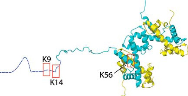
Figure 1.

Shown is the structure of histone H3/H4 tetramer with highlighted acetylation sites. Shown is the structure of histone H3/H4 tetramer (62) with boxes around the sites that were acetylated. H3 is shown in blue and H4 in yellow. H3 residues 1–20 are shown in an extended conformation as they have no discrete fold within the crystal structure. The structure was generated from Protein Data Bank code 1AOI using VMD.
