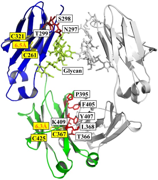
Figure 1.

Structure of Fc [PDB 3AVE (47)] presented by PyMOL. The CH2 and CH3 domains are colored by blue and green, respectively; the residues (N297, S298, and T299) in glycosylation motif in CH2 are colored dark red; the residues (T366, L368, P395, F405, Y407, and K409) involved in the interactions between two CH3 domains are colored by light red; and the oligosaccharides and native disulfides are colored by lemon and yellow, respectively.
