Figure 2.
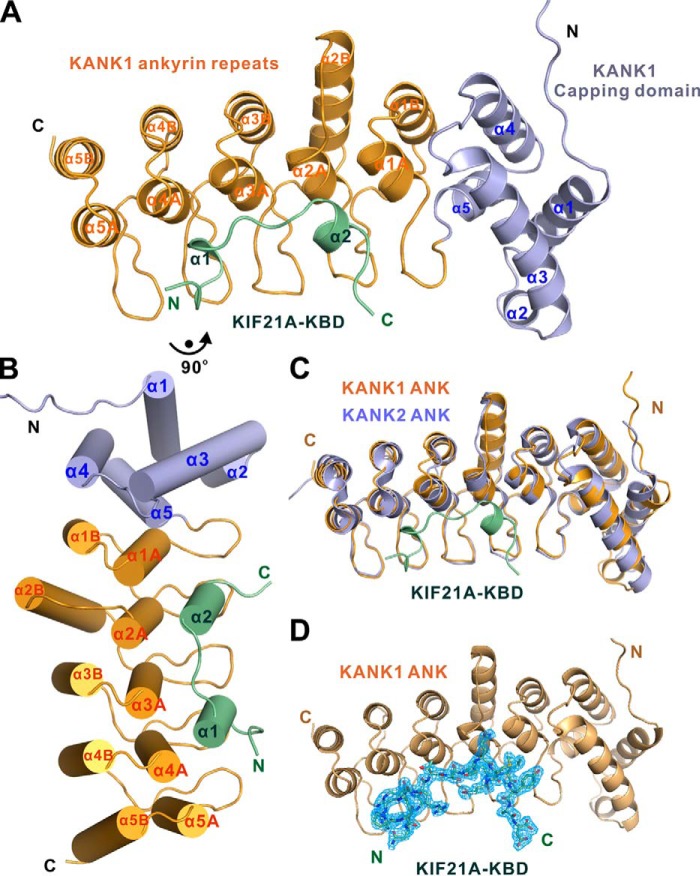
Overall structure of the KANK1·KIF21A complex. A and B, ribbon diagram (A) and cylinder (B) representations of the KANK1·KIF21A complex structure. In the drawing, the capping domain and ANK repeats of KANK1 are shown in light blue and orange, respectively. The KBD peptide of KIF21A is shown in green. C, superposition of the structures of the KANK2-ANK (PDB entry 4HBD, light blue) and the KANK1·KIF21A complex (this work). D, omit map of the KIF21A KBD peptide bound to KANK1 ANK. The map is countered at the level of 1.0 σ in PyMOL.

