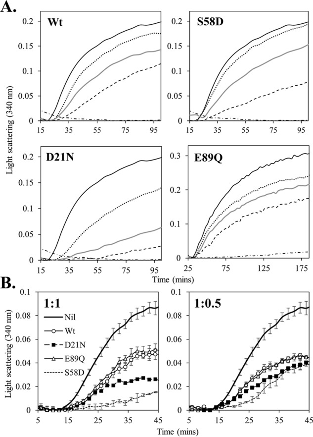
Figure 5.

Molecular chaperone activity of wild-type and mutant 14-3-3ζ proteins. A, temperature-induced aggregation (at 42 °C) of ADH was monitored by light scattering at 340 nm over time in the absence (solid line) and presence of 14-3-3 protein at 1:0.5 (dotted line), 1:1 (gray line), and 1:2 (dashed line) molar ratio of ADH/14-3-3ζ protein. The aggregation of 14-3-3ζ alone is also shown (dashed and dotted line). B, DTT-induced aggregation of insulin (at 37 °C) was monitored by light scattering at 340 nm over time in the absence (solid black line) and presence of 14-3-3 protein at a 1:1 molar ratio (left-hand panel) and 1:0.5 molar ratio (right-hand panel) of insulin/14-3-3ζ protein. Error bars, S.D. of triplicate samples.
