
Figure 1.
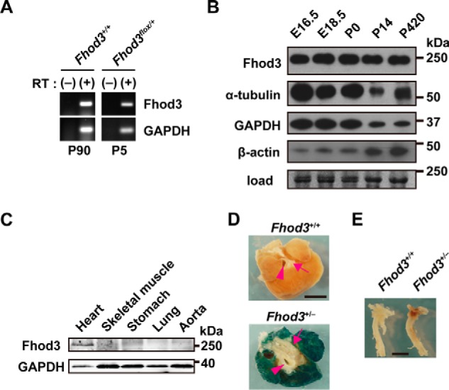
Fhod3 expression in mouse heart. A, total RNAs prepared from mouse heart at the indicated postnatal days were analyzed by reverse transcription-PCR with specific primers for Fhod3 and GAPDH. RT, reverse transcriptase. B, proteins prepared from the heart of wild-type mice at the indicated age were analyzed by immunoblot with anti-Fhod3-C20, anti-α-tubulin, anti-GAPDH, and anti-β-actin antibodies. The loading amount was verified by myosin heavy chain bands stained with fast green. Positions for marker proteins are indicated in kDa. C, proteins prepared from the indicated tissues of wild-type mice at 16 weeks of age were analyzed by immunoblot with anti-Fhod3-C20 and anti-GAPDH antibodies. Positions for marker proteins are indicated in kDa. D and E, lacZ staining of the heart (D, top view) and aorta (E, lateral view) of Fhod3+/+ and Fhod3+/− (Fhod3+/lacZ) mice at 30 weeks. Roots of aortic and pulmonary arteries were indicated by arrowheads and arrows, respectively. Scale bars, 2 mm.