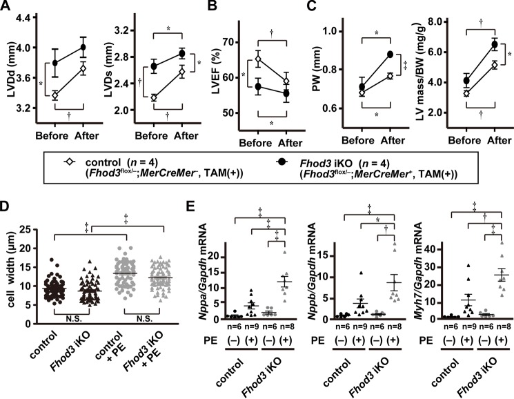
Figure 10.
Fhod3-deleted mice shows more pronounced hypertrophic response to phenylephrine infusion. A–C, echocardiography analysis of hearts of TAM-treated Fhod3 iKO and TAM-treated control littermate mice before and after PE infusion. LVDd, left ventricular dimension in diastole; LVDs, left ventricular dimension in systole; PW, end diastolic posterior wall thickness; LVEF, left ventricular ejection fraction. D, width of cardiomyocyte at the nuclei level was estimated by wheat germ agglutinin staining of left ventricle septum from TAM-treated Fhod3 iKO mice with PE infusion (n = 71 from two mice) or without PE infusion (n = 68 from one mouse) and TAM-treated control littermate mice with PE infusion (n = 67 from two mice) or without PE infusion (n = 59 from one mouse). E, quantitative real-time PCR analysis of hypertrophy-related gene expression in hearts of TAM-treated Fhod3 iKO and TAM-treated control littermate mice with and without PE infusion. Nppa, encoding ANF; Gapdh, encoding GAPDH; Nppb, encoding BNP; Myh7, encoding βMHC. *, p < 0.05; †, p < 0.01; ‡, p < 0.001; N.S., not significant.

