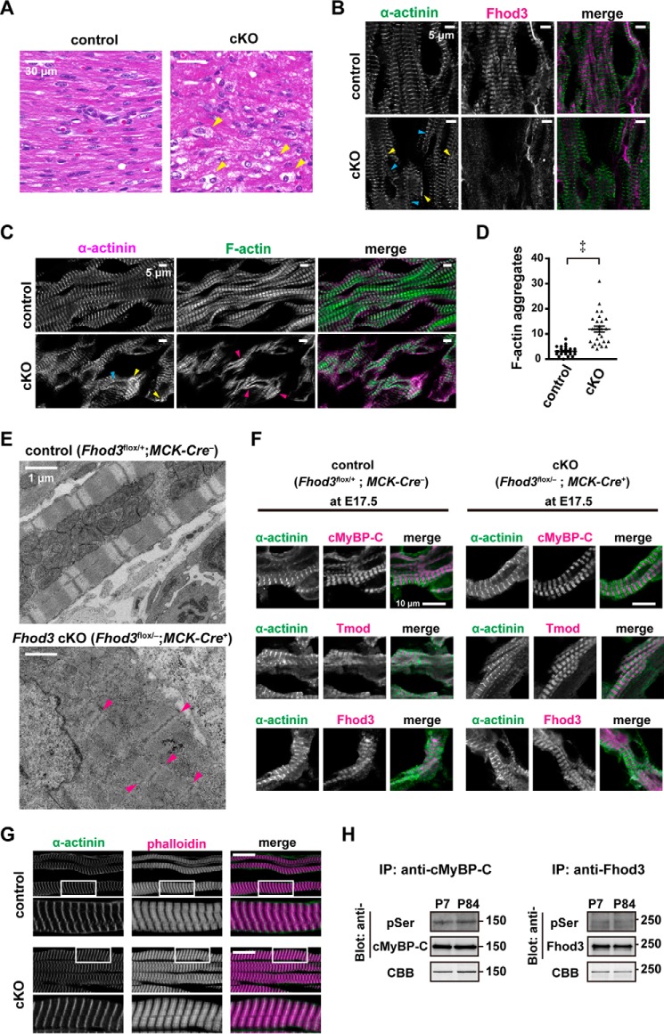
Figure 4.
Perinatal deletion of Fhod3 induces disruption of cardiac sarcomeres. A, histological analyses of Fhod3 cKO (Fhod3flox/−;MCK-Cre+) and control littermate (Fhod3flox/+;MCK-Cre−) mice at P7. Paraffin-embedded sections of neonatal hearts were stained with hematoxylin and eosin. Scale bars, 30 μm. Yellow arrowheads indicate vacuoles. B and C, confocal fluorescence micrographs of hearts of Fhod3 cKO (Fhod3flox/−;MCK-Cre+) and control littermate (Fhod3flox/+;MCK-Cre−) mice at P6. Cryosections of neonatal hearts were stained with the anti-α-actinin antibody (green) and the anti-Fhod3-(650–802) antibody (magenta) (B) or with the anti-α-actinin antibody (magenta) phalloidin (green) (C). Scale bars, 5 μm. Abnormal α-actinin signals aggregated (yellow arrowheads) or continuous along the sarcolemma (blue arrowheads) are indicated. Magenta arrowheads indicate continuous aggregates of F-actin. D, quantitative analysis of myofibrillar changes was performed by counting the number of continuous F-actin aggregates in randomly selected fields (Fhod3flox/−;MCK-Cre+, n = 28; and Fhod3flox/+;MCK-Cre−, n = 29). ‡, p < 0.001. E, electron micrographs of thin sections of hearts of Fhod3 cKO and control littermate mice at P6. Scale bars, 1 μm. These experiments have been repeated four times on three different pairs of cKO and control mice with similar results. Magenta arrowheads indicate Z lines or Z line-like structures. F, myofibrils in the embryonic heart. Confocal fluorescence micrographs of hearts of Fhod3 cKO (Fhod3flox/−;MCK-Cre+) and control littermate (Fhod3flox/+; MCK-Cre−) embryos at E17.5. Sections of embryonic hearts were stained with the anti-MyBPC antibody (magenta) and the anti-α-actinin antibody (green) (upper panels), the anti-tropomodulin1 (Tmod) antibody (magenta) and the anti-α-actinin antibody (green) (middle panels), or the anti-Fhod3-(650–802) antibody (magenta) and the anti-α-actinin antibody (green) (lower panels). Scale bars, 10 μm. G, confocal fluorescence micrographs of quadriceps muscles of Fhod3 cKO (Fhod3flox/−;MCK-Cre+) and control littermate (Fhod3flox/+; MCK-Cre−) mice at P6. Sections of quadriceps muscles were stained with the anti-α-actinin antibody (green) and phalloidin (magenta). Lower panels show the magnified views of the boxed areas in upper panels. Scale bars, 20 μm. H, in vivo phosphorylation of Fhod3. Proteins of lysates prepared from mouse heart at the indicated postnatal days were immunoprecipitated (IP) with anti-Fhod3 or anti-cMyBP-C antibodies, and the precipitants were subjected to SDS-PAGE followed by immunoblot with anti-phosphoserine antibodies or staining with Coomassie Brilliant Blue (CBB).

