
Figure 6.
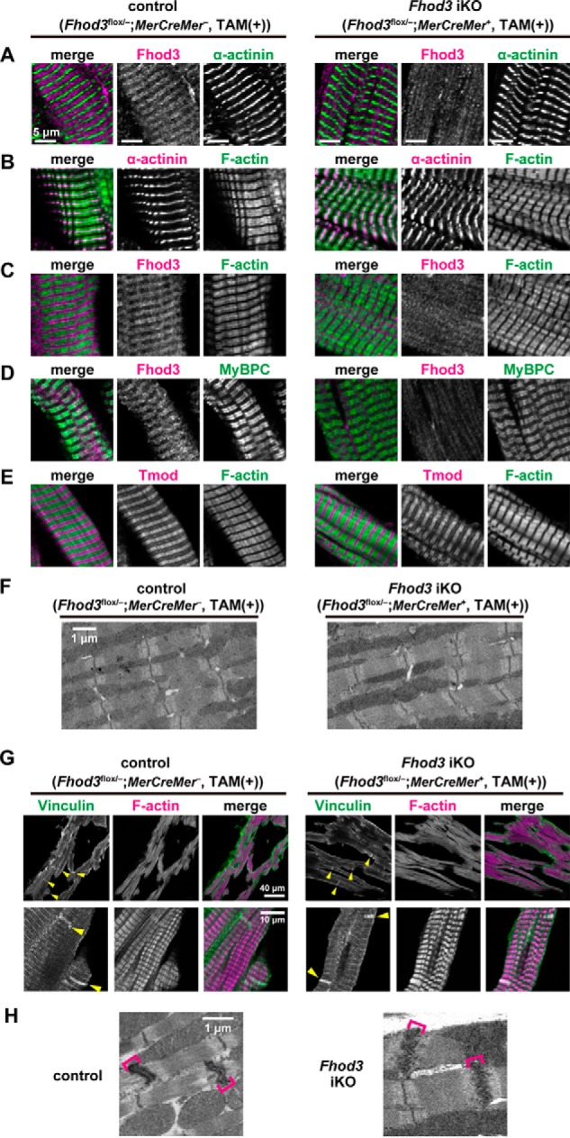
Tamoxifen-induced deletion of Fhod3 in the adult heart does not affect sarcomeric structures. A–E, confocal fluorescence micrographs of hearts of TAM-treated Fhod3 iKO and TAM-treated control littermate mice. Sections of adult hearts were stained with the following anti-Fhod3-(650–802) antibody (red) and the anti-α-actinin antibody (green) (A); anti-α-actinin antibody (red) and phalloidin (green) (B); anti-Fhod3-(650–802) antibody (red) and phalloidin (green) (C); anti-Fhod3-(650–802) antibody (magenta) and the anti-MyBPC antibody (green) (D); and anti-tropomodulin1 (Tmod) antibodies (red) and phalloidin (green) (E). Scale bars, 5 μm. F, electron micrographs of thin sections of TAM-treated Fhod3 iKO and TAM-treated control littermate mice. Bar, 1 μm. Over 10 images from each genotype were analyzed. G, intercalated discs in the heart. Sections of adult hearts were stained with the anti-vinculin antibody (green) and phalloidin (magenta). Yellow arrowheads indicate intercalated discs. Scale bars, 40 μm (upper panels) and 10 μm (lower panels). H, electron micrographs of thin sections of hearts of TAM-treated Fhod3 iKO (Fhod3flox/−;MerCreMer+) and TAM-treated control littermate (Fhod3flox/+;MCK-Cre−) mice. Brackets indicate intercalated discs. Scale bar, 1 μm.