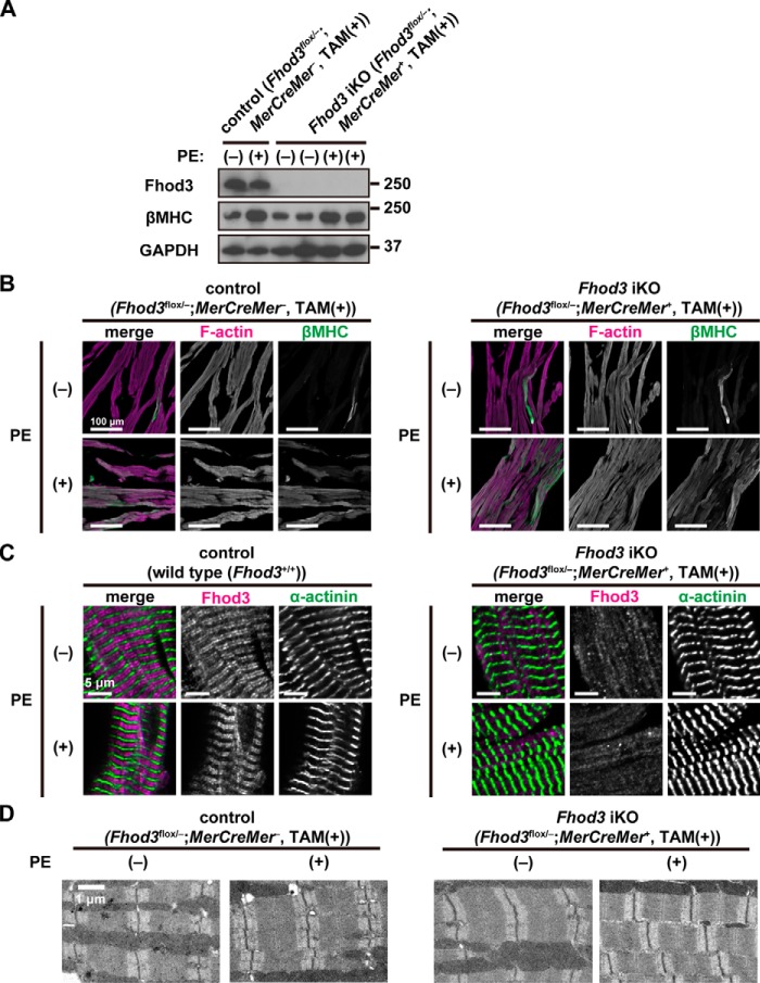
Figure 9.
Phenylephrine infusion does not induce sarcomeric changes in the Fhod3-deleted heart. A, detection of the Fhod3 protein by immunoblot analysis. TAM-treated Fhod3 iKO and TAM-treated control mice were continuously infused with PE for 2 weeks. Proteins prepared from the heart of the PE-infused mice were analyzed by immunoblot with the anti-Fhod3-C20, anti-βMHC, and anti-GAPDH antibodies. B, confocal fluorescence micrographs of hearts of TAM-treated Fhod3 iKO and TAM-treated control littermate mice after PE infusion. Sections of hearts were stained with the anti-βMHC antibody (green) and phalloidin (magenta). Scale bars, 100 μm. C, confocal fluorescence micrographs of hearts of TAM-treated Fhod3 iKO and age-matched control mice after PE infusion. Sections of embryonic hearts were stained with the anti-α-actinin antibody (green) and the anti-Fhod3-(650–802) antibody (magenta). Scale bars, 5 μm. These experiments have been repeated three times on two different pairs of iKO and control mice with similar results. D, electron micrographs of thin sections of hearts of TAM-treated Fhod3 iKO and TAM-treated control littermate mice after PE infusion. Bar, 1 μm. Over 10 images from each genotype and treatment were analyzed.

