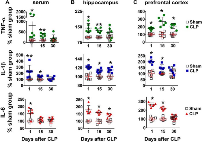
Figure 1.
Content of pro-inflammatory cytokines in serum, hippocampus, and prefrontal cortex at 1, 15, and 30 days after CLP. The content of pro-inflammatory cytokines IL-1β, IL-6, and TNF-α was assessed by ELISA in serum (A), hippocampus (B), and prefrontal cortex (C). Values represent relative quantification considering control (sham group) as 100%. Scattered individual data points (n = 6) and standard deviations are represented. Differences between sham and CLP groups on each day were considered significant when p < 0.05 according Student's t test (two-tailed) analysis (*, p < 0.05, and **, p < 0.001).

