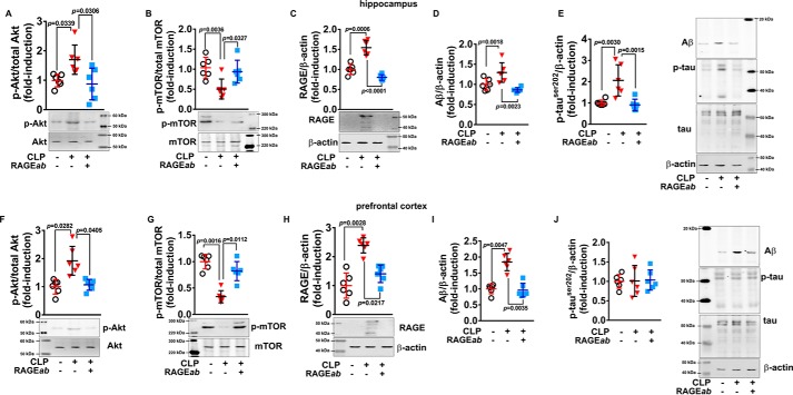
Figure 11.
Akt and mTOR phosphorylation are associated with RAGE and markers of neurodegeneration in brain. RAGEab was administered bilaterally into the hippocampus at 100 μg/kg at days 15, 17, and 19 after CLP. Hippocampus was isolated, and the levels of phosphorylated Akt (A) and mTOR (B), RAGE (C), Aβ (D), and phosphorylated Tau (E) 30 days after CLP were analyzed by Western blotting. Similarly, in isolated prefrontal cortex, phosphorylated Akt (F) and mTOR (G), RAGE (H), Aβ (I), and phosphorylated Tau (J) were assessed. Scattered individual data points (n = 6) and standard deviation are represented for all data. Representative Western blottings are demonstrated. Differences between groups were considered significant when p < 0.05 according to one-way ANOVA with Tukey's post hoc test; individual p values are depicted.

