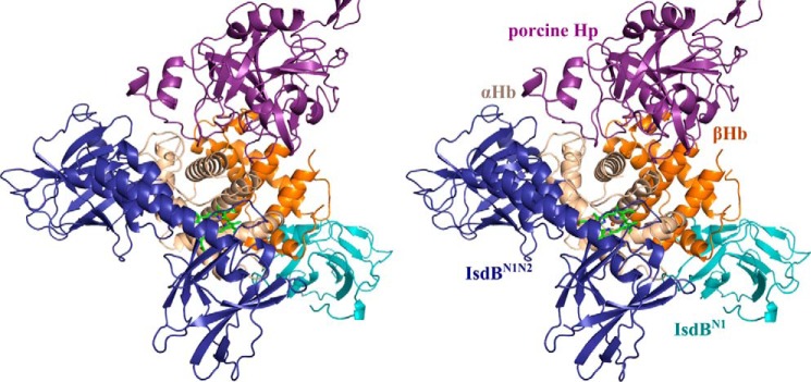
Figure 10.
A stereo overlay of the Hp·Hb structure with the IsdBN1N2·Hb structure. Shown is superposition of porcine Hp·Hb (PDB code 4F4O) with one of the α/β Hb dimers of the IsdBN1N2·Hb structure. Hp binds at a distinct site from IsdB. Hp is shown in purple, and IsdBN1N2 and IsdBN1 are shown in dark blue and light blue, respectively. The αΗβ and βHb subunits are shown in beige and orange, respectively, with the αHb heme shown as bright green sticks.

