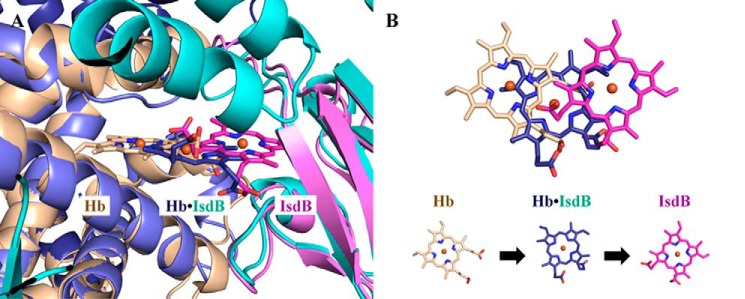
Figure 9.
A model of the heme extraction pathway. A, heme positions observed in αHb, the IsdBN1N2·Hb complex, and isolated IsdB. The αHb chain of oxyHb (PDB code 2DN1; beige) overlaid on top of the complex αHb (dark blue) is used to represent the pretransfer heme position in uncomplexed, folded Hb. The heme-bound form of IsdBN2 (PDB code 3RTL heme conformation A; pink) overlaid on top of the complex IsdBN2 domain (cyan) represents the completed heme transfer reaction. B, top-down view of the positional changes of the heme molecule shown in A. The heme iron in the complex structure (dark blue) is ∼5 Å away from both the initial and final heme iron positions.

