
Figure 5.
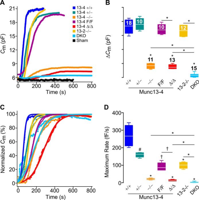
Munc13-4 regulates the amount and rate of MC exocytosis. Shown are capacitance (Cm) recordings from peritoneal MCs during exocytosis stimulated by intracellular dialysis of GTPγS and Ca2+. Access to the cell interior under the whole-cell patch clamp recording technique was achieved at time = 0. Sham, Munc13-4+/+ MCs dialyzed with a solution lacking GTPγS. DKO, double Munc13-2 and Munc13-4 KO mouse. Color legend in A applies to all panels. A, representative traces of cumulative Cm. B, total Cm change (ΔCm) above baseline. n = number inside boxes, applies to B and D. C, representative normalized Cm traces. D, maximum rate of ΔCm (rate between 40 and 60% of total ΔCm). Line, mean; box, 25th–75th percentile; whiskers, 5th–95th percentile. # = p < 0.05, † = p < 0.01, * = p < 0.001; all comparisons are to Munc13-4+/+ unless otherwise indicated.