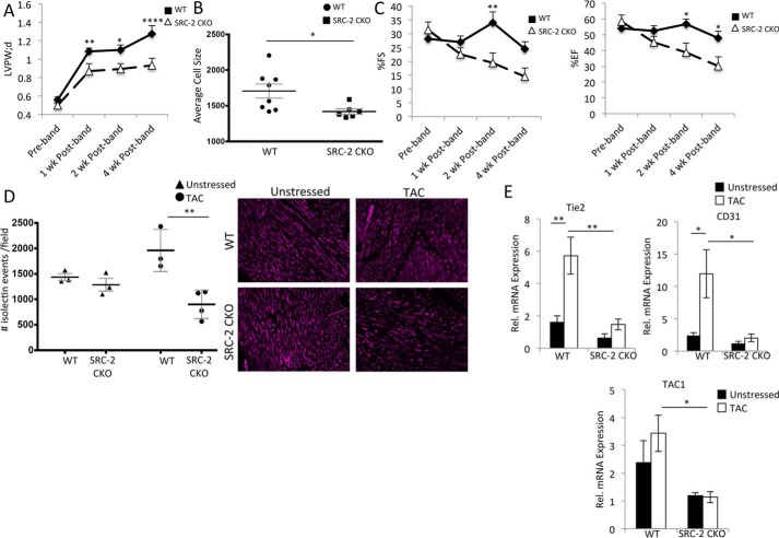
Figure 1.
SRC-2 mice have decreased vasculature post-TAC. A, left ventricular posterior wall thickness (LVPW) echocardiography measurements for WT and SRC-2 CKO mice 4 weeks post-TAC. n = 7–15. B, heart sections from WT and SRC-2 CKO mice were stained with wheat germ agglutinin (WGA), and a cross-sectional area of cardiomyocytes was measured. n = 8–12, ≥400 cells/mouse. C, percentage ejection fraction and fractional shortening derived from echocardiography as described in A. D, isolectin staining of heart sections as in B. Number of vessels per microscope field is graphed. n = 3 mice with 4 sections/mouse analyzed. E, qPCR was performed for the indicated genes on total heart RNA from WT and SRC-2 CKO hearts before and 4 weeks after TAC. Rpl32 is used as an internal control, and data are presented relative to unstressed WT. n = 4. *, p ≤ 0.05; **, p ≤ 0.01; ****, p ≤ 0.0001. Tie2, tyrosine kinase with immunoglobulin-like and EGF-like domains 2; TAC1, tachykinin precursor-1. Error bars, S.E.

